Introduction & Research Overview — Chunyue Yin, PHD
I’m Chunyue Yin. I’m an associate professor, in the division of gastroenterology, hepatology and Nutrition at Cincinnati Children’s Hospital, and I’m also co-director of the Center for Undiagnosed and rare Liver Disease. And I will be the moderator for this session and Emily actually give me 10 minutes to talk. So I will actually share stories with you guys, because, You rarely hear researchers share stories.
Feel very um fortunate to do my job because uh when you talk to most of the other researchers, they usually spend their whole career in the lab um studying cells, animal models, and DNA um but they never get the opportunity to meet a patient who has the disease they are studying. But in my position, I actually get to meet you guys and then I work alongside with uh physicians, pathologists, human geneticists, industry partners, so we, especially families and patients, so we all work together and I get to see research not just as an experiments, but, researchers, put a face on the research is a family, research is a story. So in the past year, I actually, got to meet two families who lost their kids over 50 years ago, due to unknown cholestasis. At that time, liver transplant was not available and there was no genetic testing.
For the over half century, these families just live in this constant worry. They didn’t know what happened to their kids. They were worried, if this would happen again to them, how about their, other kids and grandkids. And then as a mom, I understand mom always feel like, did I do something wrong, cause this.
So with the new technology in sequencing, and new knowledge in biology, we actually got to revisit these cases. We successfully retrieved DNA from, samples they preserved over 50 years ago. And then we sequenced the DNA we actually were able to provide a diagnosis for both families. It’s, it’s that type of story make you feel like make me feel the first time like my research matter, my research is helping people and change their lives.
They got their closure. Then more recently, I met another family who also lost their baby boy several years ago. At that time, whole exome sequencing was already available. They went through the sequencing, but, they didn’t find any, mutations in the genes that we know to cause cholestasis.
So we decided to revisit the case because of the new technology. And then uh we actually found changes in the gene that had never been associated with cholestasis before, but because of the knowledge we gained over the years, we thought this could be a candidate for their uh disease. So in my lab we made zebra fish, carry changes in the same gene and then uh we found the fish actually developed liver disease that was similar to the patients. So we currently are studying the fish mutants.
And then try to understand the mechanisms so hopefully we could find a cure, therapeutic for future patients with this uh mutation in the same gene and also uh we can improve diagnosis for these patients. So I share these stories to let you get a feel how research have evolved and how research have um changed everybody’s life and also my life. I feel I have the best job in the world. Also it’s important to, for you to realize that um.
Research start with family, you have a met needs, you have a question, and then we turn it into a research project and we all work together, families, advocacy group, researchers, clinicians, and also, industry, we all work together to move this research forward. So today, uh we have 3 world-class experts in Pfake research, who are going to give you updates on, diagnosis and treatments, so you can see, where the future holds. So
New Developments in Genetic Testing & Diagnosis — Richard Thompson, MD PHD
Thank you and you, if you’ve got the best job in the world, I’ve definitely got the best job in the world. I have said that many times before. I’m super lucky that I can see patients, go to the lab, try and work out what’s wrong with the patients, try and work out how we can try and make them better, and then we are getting somewhere now and we are starting to get better treatments, which is amazing. So I was commissioned to talk about future advances or recent advances in diagnosis, and you heard a lot from Paula earlier on today, and I’m going to touch on some of the same topics and expand that.
But I’m gonna start more fundamentally about the concept of diagnosis, to be honest, and you heard from several people and very eloquently from Stuart earlier today about how it took several decades for him to have a diagnosis. But you’ve also heard um from various people that er not everyone with a disease behaves in the same way. So I would argue that actually having a diagnosis isn’t really the most important thing, and in fact it may not be the right thing to do to anyone. What I believe that we’re all trying to do is understand what’s wrong with someone, and try and explain uh why, what is likely to happen to them in the future, and what we can do to improve that.
OK? And I don’t think that actually giving someone a label is necessarily the best thing to do. I think we have to understand each individual’s biology. And then address them as individuals rather than giving a label and assuming they’ll behave the same way as everyone else that we’ve given the same label to.
So you’ve heard this concept of phenotypes earlier on today, and I believe that really the best way to talk about someone’s disease is not giving them a label, is to describe the phenotypes, the different components of the disease that they are manifesting, whether that’s based on observation, what they’re telling you, or blood tests or scans, but they all constitute the phenotypes, the parts of the disease. And those phenotypes that we all have, we’ve all got variation in our characteristics to some extent or other can be explained by genetic variation, and that is really what we’re trying to do with genetics now. So to go about how we, how we have been doing that, and you’ve heard a little bit of this already from Paula, is we’ve got this quite rapidly advancing technologies which have allowed us to make progress and you heard from Ron earlier on that there was the three of the three biggest, genes in PFIC were all discovered in the same year in 1998, and that’s not by coincidence. It’s, it’s, it’s partly led by technology and the rapid advances in sequencing that was happening then, and that was what we now call Sanger sequencing.
But since then there’s been further massive advances in sequencing which you’ve heard something about and I’ll go into in more detail, which have allowed these panel sequencing, exome sequencing and genome sequencing. Actually sequencing the genes is only part of the story, as I think you’ve already heard, is we’ve got to then classify the variants that we find in two ways to the likelihood of effect because what you see on reports, generally on genetic reports is how likely variants are to have an effect on the protein. But we also now have to begin to understand the degree to which variants might have an effect on the protein, not just whether they do or they do not. And then understand how that particular variant in that particular individual has contributed to the phenotype which they’re presenting with.
And then understand, er, on the basis of the genetics, if we can, the likelihood of response to treatment. So to go back to the beginning, so as you know, genes are contained in DNA and they’re packaged into chromosomes. And in humans we’ve got 3 billion bases of DNA. So each of those letters, A, C, G’s and T’s, there’s 3 billion, in our genome.
So we’ve inherited two copies of those 3 billion bases, one from each of our parents basically, and they’re arranged in these 23 chromosomes. And we have about 20,000 individual genes, and the majority of that is what we call non-coding DNA, so only 2 or 3% of the DNA is actually coding for the bits of the genes that actually make the proteins. We realize that some of the rest of it is regulating how those er genes are used and the proteins are made, er but to be quite honest, the majority of the DNA we do not understand yet er what it’s er relevance is to being healthy, never mind disease. I’m sure we will unravel that over the next few years, but the majority of the information we have about genetic variation.
And contribution to disease is just focused so far on those few percent of DNA which we understand its relevance because it’s, it’s there to make proteins. And as you’ve already heard um from Paula. The DNA that’s the template that we’ve inherited from both of our parents for an individual gene is used to make an RNA copy, and then the RNA copy is used to make the protein. And just to explain some of the technologies, I wanted to show you this, which is, this is an example of a gene in DNA at the top and to point out the actual gene is broken up into individual exons.
So the bits in the spare boxes, the number exons, are the parts of the gene that we understand the best because they’re the bits of the gene that are used to make an individual protein. From that DNA we make an mRNA and initially the mRNA is like this. Or the RNA is like this is a pre-mRNA. It contains the boxes, but it also contains the bits in between which we call introns, which are not relevant to the code to make the proteins.
And they are called, that is spliced, the RNA is spliced until we get rid of all those intervening sequences and we just have the exons which have the code for the proteins, which is what we use then in individual cells to make proteins. And I’m showing you this because this becomes important because um as I’ve said already, we understand variants which are in the bottom section there in the exons that make proteins and the missense and truncating mutations you’ve heard already are ones that occur in that coding sequence. But we now realize there’s variants occurring outside that sequence that are in the DNA but they may be in the gaps in between the introns which are in the between the exons which are contributing to the disease, or in the upstream regulatory parts of the DNA which are not which are turning the gene on and making it be used in the appropriate time in the appropriate cells. So we’re only just beginning to explore variation which isn’t outside the coding sequence, which is much easier to get your head around.
So you’ve seen a slide very similar to this from Paula, and at the top of the missense changes, and these are the easy ones, this is where the code is changed and you make a different amino acid to the one that you’re supposed to make, and sometimes that has a minor feature, minor consequence, sometimes it has a severe consequence. Those are the missense changes at the top. And you’ve also heard about the stop mutations, which are the nonsense mutations, which is where there’s a change in the sequence, which means that the protein is stopped at that point and you don’t make a full length protein, and that usually has a severe consequence. What I’ve just been talking about is splicing because you’ve got to splice these bits of different exons together, and there are now a growing number of variants in the very close to those axons or actually further out in the introns which change the ability of the RNA to splice, and if it doesn’t splice properly into that continuous sequence, you can’t make normal protein.
As I’ve already told you, upstream, further away from the gene are regions which are essential to telling the cell to use that gene, we call these regulatory elements and promoters which turn a gene on or turn a gene off in a particular cell at a particular time. And for some genes now we’ve got a good understanding of the regulatory elements and many others we don’t. Then we’ve got more complicated um types of changes to genes, some of which I’ll give you examples of and I think are now emerging as important disease genes. The first one, small insertions and deletions, are actually more easy to understand.
That’s rather than being a change in the code, there’s actually an insertion or deletion from the code, so one or more nucleotides, these bases, the ACG’s and T’s may be missing or there may be some extra ones put in, and that really disrupts the sequence. But we can, we now realize that there’s many changes which are larger insertions or deletions where there’s a major chunk of DNA has been chopped out or is inserted, and they are now much easier to detect than they were in the they were previously. And I’ll show you how we can do that. And there’s even more uh complicated genomic rearrangements where there’s actually bits of DNA moved from one part of the genome to another part of the genome which can completely disrupt a gene.
Or it can even move a whole gene to another part of the genome which has consequences. So the sly site changes, the very close ones, very close to the axons we’ve been picking up for many years, but the ones further away are the ones we’re missing until recently. The promoter changes are the ones we’ve been missing until recently, and these large insertions and deletions, the technology is improving to detect those as well. So Sanger sequencing, which is what we were all using back in 1998 when we discovered these genes, was, it was a massive breakthrough, but it was painful.
We had to amplify each of those exons individually, and for each gene, and then sequence those, individual sections of the gene. So we could only look at one patient, we had to amplify each exon separately. We could only sequence in one direction, and then the output you got, and I’ll show you a picture in a second. It was a mixture of the DNA because, as I say, we’ve all inherited two copies of every gene, one from each parent, and very often we, if we get a change, we’ve got it from one parent and not from the other.
And so what we look at when we’re seeing it is the DNA is the mixture of those 22 basically two copies that we both have. And that can make it very difficult to distinguish exactly what’s going on and I’ll show you an example. But these are other examples of why, Sanger sequencing, Is just sequencing a mixture of whatever’s there, and I’ll show you why that’s important, it’s it’s very difficult to, pick up on here, I’m sorry, but this is what sanger sequencing looks like, it’s a series of um AC’s, G’s and T’s, and in here, if you look here, there’s a black and a green peak superimposed, and so that’s a person who’s heterozygous, there’s a single base change. And it’s quite difficult to see, but you can see that in this case it has come from the father who’s got it in the same position in the middle who’s got a black and a green peak, whereas the mother at the bottom has only got a black peak.
And so what you are seeing though in the patient, and that’s why I’m showing you this, is that the DNA is a mixture of the two types of DNA, one from the father, one from the mother. However, that’s Sanger sequencing and that’s largely been replaced for discovery. We still use it often for confirmation of changes or if we want to look very quickly to look at the mutations that are in a child, we may just use the DNA from the parents just to confirm that. But for gene for discovering new patients, er, we now use all this NGS technology, whether it’s panel sequencing, exome sequencing, or whole genome sequencing.
It is essentially the same technology. If you take, this is supposed to represent a long strand of DNA, a whole chromosome, many millions of bases of DNA, it actually gets chopped up into short fragments, typically a few 100 base pairs out of that 3 billion base pairs. These are just a few 100 base pairs of DNA. And they have modifiers attached to the ends, and the modifiers allow us to do the sequencing, but they also have a code which tells us which patient it’s come from as well, because in a minute we’re gonna mix all the DNA together from many patients into the same tube, which was a horrifying thing when we started doing it.
But we’re gonna put this code on such that when we actually get the data out, we can look at the code and it’ll tell us which patients it’s come from, so we, it’s, we actually do many patients in the same tube. What we do with those fragments of DNA is we put them into a machine and individual fragments will stick to one of these little red and blue, they’re not really red and blue obviously, but, on here they look like they’re red and blue on the surface of a sheet of glass, and then we make multiple copies from each of those little bits of DNA, so they’re all identical copies. You end up with a cluster of identical bits of DNA on a glass plate, and that is what is actually sequenced. And at the end of that process, this is what the data looks like.
And this is a real patient, um and this is an example from um from BSEP. And um what we’re seeing at the top, if you go and look at the top, there’s the blue rectangles are individual exons of this gene. And the narrower blue line with the arrows are the introns in between the exons. And what we’re seeing down here are lots of short segments which are individually gray, and these are what we call reeds, and each reed is about 150 base pairs long.
It’s been generated by sequencing one of those clusters, and all those millions and millions of reeds have then been mapped against the normal sequence. So the computer doesn’t know where they’ve come from, it just takes all these reeds, it looks at the sequence and it says, oh, that’s come from. That bit of Exxon of that Exxon from that bit of that gene. The computer does all that for us, and so we get these literally 100s of copies of the sequence from each gene.
And as you can see here critically, we’re only getting any of these reads where there are the blue axons at the top. This is actually an example of exon sequencing, a whole exome sequencing, so we’ve sequenced all the exons, all the blue bits if you like, of all the genes. But if we had the same data from a panel looking at BSEP, it would look exactly the same. And critically, what we haven’t got is any data, any reads from the bits in between the blue because we haven’t interrogated those bits.
Now if we zoom in on one of those regions, it looks like this. And so at the top we’ve still got, if you look on the second line, there’s a blue line across there, which is the axon on the right hand half, and then it goes into an intron on the left hand half. And we’ve got, what we’ve got below here, and this is just a very few of the hundreds of reeds, that every one of those little colored lines has an arrow at one end. Guess they’re going in one direction or the other, but they’ve all been stacked up by the computer.
And looking at that, it’s very difficult to see, but what you can see is that most of them is the same color all the way down, and they’re completely normal. If I then magically take away all the ones which are normal. You will see it’s in the middle. There is what, it should be a T in half of those reads is a C.
And so this is an example on next generation sequencing, whether it’s NGS panels or exomes or WGS. This is what the real data looks like, is that you can see whether it’s going blue in that direction or red in that direction doesn’t make any difference. But in half of those reads, there’s a C at that position, half of them, it’s a T at that position, and this is what the raw data looks like from any form of NGS sequencing. So NGS can be uh targeted or it can be the whole genome.
So literally the whole genome is the easiest to understand, even though that seems like it’s the most recent technology, and it’s technology which has been used for research but is increasingly used for diagnostics, where we literally sequence the whole of the gene, the whole of the whole of the genome. So that DNA I showed you at the beginning and the swirls of DNA, we literally just chop it up, make it all into those fragments with the tags on the end, and we sequence every single little bit of it. However, what we’ve been doing for the panel sequencing and for exosome sequencing is we’re then using a tech a technology to fish out the bits that are relevant to the bits that we’re interested in. And we’ve done that for various reasons in the past because actually it’s technically, it means we’re sequencing much less of the genome, we end up with less data and we have fewer, they’re less information from genes that we’re less interested in and we can focus on the genes that we’re interested in.
But the truth is that doing the whole genome sequence where we literally sequence all of it actually gives us more even coverage, so technically it’s better. And it gives us information about the intervening sequences, although at the moment we’re much less able to understand what all that means. So basically the technology for these uh different forms of next generation sequencing are very sim are all very similar. The reeds, as I’ve shown you are aligned to the genome, all those gray lines that become colored lines are just stuck to where they should come from.
And then as I’ve shown you, the variants are detected. And then there’s quality filters, etc. And then we rely on a computer to give us as much information as possible to see what the likelihood is that those variants are doing anything and what we know about them. One of the limitations of the technology we’re using at the moment is that all these little fragments are just short fragments.
And it’s very difficult to, the computer puts them onto the genome where it thinks they’ve come from, but it’s not always right. And if the genome’s been rearranged, it will not work out where the rearrangements have happened because it’s only interrogating very short segments of the genome. So if there’s 2 changes and many of the patients in the room have got 2 variants, one from their mother, one from their father, and when we look at it by NGS, we can’t tell that those 2 changes have come from one from the mother and one from the father, cause there’s just 2 changes, and we, because of the fragments are short, we can’t tell that they’re 1 from each parent, and it’s only by looking at the parents that we can then tell that one’s come from one and one’s come from the other. There are new technologies which are use much longer reads of DNA.
We can use millions of base pairs of DNA as one continuous read, and that will allow us in the future to establish the inheritance in a single experiment. The newer technologies also mean that we’ve got, we’re better at measuring what I call copy number variants or CMBs, which is where a whole bit of DNA has been duplicated or been deleted. And, this is the sort of technology that’s allowing the long reach sequence. This is a, Oxford nanopore sequencer.
This is a 48 cell. I’ve got a 96 well cell in the lab now. These are amazing bits of kit, they will read millions of bases of DNA continuously. They give you the information in real time, so one thing we’re using them for is for brain tumors, and the surgeons sample the tumor.
We put it straight on the machine. It requires almost no preparation, and it starts generating data in real time. We don’t have to wait for the experiment to finish, and usually within, literally half an hour, 1 hour, we’ve got data coming out, and in real time the people can classify the tumors and decide how malignant it is and what sort of surgery they need to carry out. But these, that the equipment that we’ve got will now do 32 whole genomes overnight.
That’s the sort of technology that we’re now moving to, which is quite extraordinary. Just to give you another example of what I’m trying to get across that we are now able to detect, which we couldn’t previously, again we’ve gone back to a real patient with BSEP deficiency, the grays or the um reeds against those four exons at the top. Well, the sort of thing that can happen is that you can get that exxon there gets duplicated, so we get two copies of the same axon, and that’s gonna cause major disruption to the gene. But if we did that by Sanger sequencing, we wouldn’t actually detect that at all.
If we did that by sort of early next generation sequencing, we wouldn’t detect that either. But the current next generation sequencing, whether it’s panels, genomes or exomes, would now detect the fact that we’ve got this two copies of an exon here, which would in many cases, grossly disrupt the function of the gene. If we go back to the normal sequence here, as I say, if there’s a change in the middle of this intron in between the two axons, it’s not changing the code, but it could be affecting the way the code is used, the way the exons are put together. The sequencing at the moment, whether it’s panel or exomes, is not going to detect that because there’s no gray, lines underneath that.
But it’s only by doing a whole genome sequencing that we’re now discovering many variants which are deep into these introns. But at the moment, the truth is we’re much less able to predict what the consequences are. But I don’t think there’s any doubt, that we will over the next few years discover more and more of the disease causing variants are deep into these introns. OK, I think I’ve more or less said what these amount to, but the exomes are just panels which cover all the genes.
Panels are just reduced ones where we just get the genes of interest. The advantage of doing an exome is that if you discover, like Cheng Yu has just said, if we discover a new gene, then it’s already been sequenced and we can look at that if it’s already on an exome. But the risk of exomes and genomes is we find variants in other genes which are not relevant to the disease, which are incidental findings, which may be clinically important to people, but are not part of the question that we’re asking at that time and could have implications, and that’s a whole different, kettle of fish. So the exomes do allow us to look at new genes quickly, but the genomes give us actually much better coverage, definitely allow us to look at rearrangements in the genome and will allow us to look at intron variants.
The classification that you see on reports is between, I’m running out of time. The classification that you see on reports says that something’s pathogenic, likely pathogenic, uncertain, likely benign. That’s been constructed by using these 26 variables about a variant. And then using a scoring system on the right hand side to decide whether they’re pathogenic, likely pathogenic or not.
And as I said right at the beginning, this is, you know, this is the this is the state of the art, but it’s also only telling us about likelihood, it’s not telling us about severity. I’m gonna have to move on quickly. That’s more or less the same thing. But what we really want to know is the relationship between the function and the phenotype.
And I’m not going to go through these in details, but this is the way my brain thinks about it. The X axis, the horizontal axis is the amount of function. The Y axis is the chances of being disease free. We’re all hoping up at the top right-hand corner.
As I said earlier on in the questions, I think that most of the patients we discovered in these genes were at the bottom of the left-hand corner where they had very little function early onset severe disease. But we’re now establishing the relationship, more subtle relationship between people who’ve got varying degrees of loss of function, not only the severity of their disease, but critically their likelihood of response to treatment. And most of the, most of the genes we’ve been talking about, today, and most of the genes we’re interested in do behave largely like recessives and got a curve that’s similar to that one at the top, whether it’s BSEP or FIC1. But critically, MDR3 is the one that’s different, and this is why we’re seeing heterozygotes who are in the middle where the blue dotted line is.
They haven’t got the severe disease that the kids had that were originally diagnosed with MDR-3, but they have got a risk of disease, but it’s a hugely more complicated relationship. So I’m gonna wrap up by saying that you’ve already heard about using clinic about genetic counseling, but I do believe that once we’ve got the genetic data, we then have to go back to the clinical data, look at the histology if we’ve got it, get the input from clinical genetics and the laboratory genetics to try and put together. The information to decide what those variants are contributing to the patient’s phenotype. And to push this further forward, we’re gonna need data on patient outcomes, which NAPA is one of the main tools that we’re using for that to look at the relationship between er disease and response to treatment.
But also we’re gonna have to do more laboratory data, particularly for some of these intronic variants, which some of which are undoubtedly disease causing, but at the moment we cannot classify. So I hope I’ve convinced you in a little bit that really making a diagnosis isn’t the most important thing. What we need to do is understand the genetic contribution in an individual to their phenotype, so that we can then try and give some idea about prognosis, we can try and predict complications along the way, we can ideally tailor treatment and predict response to treatment and eventually predict adverse effects of treatment and hopefully in the end, and I admit we’re not there yet, individualized therapy for each individual patient. Got there in the end.
OK.
Disease Mechanisms & Gene Therapy Progress — Henkjan Verkade, MD PHD
Uh thank you all for Given the chance to speak to you again and uh it’s my pleasure and I have an experiment today. I know we’re talking just before lunch, which is quite difficult, well, speaking after lunch is more difficult, but before lunch is difficult. I have an experiment and I need some time to get you trained. In the end, I’ll show you the very recent result from a month ago on PFIC 2 BSEP deficiency treatment in mice.
I know. We are medical doctors, we are not veterinarians, but still, this is the state of the art of research, and I would like you to understand it. I will show you the abstract. That’s how it works, the abstract of the paper, and we go through it together and the way to it, towards it, would like to train you to understand what they’re actually doing and how this works out, probably for the future of the disease.
So be with me. Here we go. This is the disclosure of the different uh it’s good to know for this. Audience, 6 out of 7 of these companies that, that I have contact with, and I do things with, 6 out of 7 is working at different stages on treatment of rare liver diseases and usually cholestatic liver diseases, and there are companies that you have never heard of because they’re still Pipeline, they’re still biotech, they’re still not, they don’t have the product yet, but they’re working on it.
So that’s also perhaps good to realize. So I would like to uh Tell you in 3, these 4 topics, back to the basics, pilots and beyond. 2 forms of choostasis, how and where can we intervene in choostasis, and then The real proof of the pudding, the example of novel treatment of PFIC in mice. So back to the basics, and Dr.
Ron Sokel already also alluded to it. Bile acids in general, for, for this occasion in general, have two functions. Biolacids are actually soaps. Soaps like detergent.
Soaps are the way to dissolve fat or fatty substances in a water environment. And like a soap is even if you do your dishes, you need a soap. Soap is then foaming. And after you’ve done the dishes, the foam is gone.
Usually the soap is still in the water. But it’s then saturated with fat, and hopefully the fat is from your dishes into the water with the soap and then you then you have cleansed, cleaned your dishes. And to the functions, or major functions is actually to get rid of hydrophobic substances, lipids from the body, like for example, herbs. Can I go back? Yes.
Like, cholesterol, plant sterols, conjugated bilirubin is, on drugs. We have many drugs that you secrete into bile. And whereas it needs secretion into bile, when it, the bile enters the intestine, it’s the reverse, you need the solubilization to make it more soluble, the fat to be taken up. So it has a dual function.
And that’s, for example, fat diet, as well as vitamins, fat soluble vitamins. In gray, you see, we have all the functions of bile acids we have realized since about 10 years in glucose homeostasis, in metabolism, in. But I will not concentrate on those because those are not the major problem in PFIC-type diseases. So this is the scheme of enterohepatic circulation.
Enterohepatic circulation, entero is intestine, hepatic is liver. And this is, it shows the cir it shows the circle between on the upper triangle of the liver, and then on the lower right corner of the intestine, and it circles around because bile acids are kind of precious molecules. It takes the body 38 enzymatic steps and and a lot of energy. To make them uh out of just from scratch, from cholesterol, it’s a little bit less, but still, they’re precious energetically and the body wants to conserve them.
So this is the way and you can see if you do not have bile acids. And this is an experiment we did a long time ago, about 25 years ago. This is in rats. If you put rats, you give them a bile stoma.
So you take out all the bile that is secreted from the liver, and you put it out of the body, like an enterostomy, kind of like a, like a biostomy. But you don’t have cholestasis. It’s, it just fluxes, but it doesn’t get into the intestine. And then you can see on the left side.
If you, the mice are just on a regular, the rats in this case, red diet, which is a kind of chow, the amount of fat in the stools goes up about tenfold. This is 1 g per day. Whereas in normal with intact circulation, it’s only 0.1 per day. So 10-fold up.
And on the right side, It shows that dietary fat absorption in us also, as in rats, it’s very efficient if you don’t have any disease. It’s about 95 to 97% of all the fat that you ingest, you do absorb. If you don’t have completely don’t have bile acids in the intestine, and you have a low fat diet, it’s still pretty efficient. It only goes down to 89%.
However, if you push the system and you eat a very fatty diet and high fat diet, then The balance is gone because you don’t have capacity to compensate. And then your fat absorption goes down to about 50%. So this enterohepatic circulation. Functions and I like the the way it was described this morning also by the slide in the pool.
This is actually the circulation. Works only based on pumps. And that’s also if you, I think many of you have ever gone on a slide. I’m not talking about the person now, but on top of the slide.
It’s not a dry. It’s not a dry slide. There’s some water there. There’s some water flowing to make you go.
Well, this is based on pumps. If you don’t have the pumps, the slide would not work, because it would be very difficult to slide. These are 3 pumps with very romantic names. The first one is BSEP, that’s from the liver towards the bile.
Then you have IBT from the intestine back into the system. And you have NTCP from the blood into the liver. Those, we have more transporters, but those three are characteristic, and those are important for the ropetic circulation, particularly for this talk. And to make it a little bit more real life, but still schematic.
BSEP on the top right is transporting conjugated bile acids. It was already said we have bile acids, but usually bile acids are linked to an amino acid. And amino acids is, is a building block of proteins. Well, two specific building blocks in human are linked to biolacid, taurine or glycine.
And they are depicted by the yellow small circle, and the bio acid itself is the green bigger circle. They’re transported to BSEP uh to the bio. And then the end of the small intestine, I bet, I will not go for the details of the names, but I bet. It’s kind of a vacuum cleaner.
It sucks out most of the bile acids from the intestinal lumen back into the system, where NTCP. Brings them back to the liver to be secreted again to make the circle go round. This is not the whole story, however. Because part of the bile acids are not.
Vacuum cleaned by the intestine by iPad. It originally it was thought this is about 9 95% is taken up by IBET, but that’s not, that’s an overestimation. It’s rather about 80%. And about 20% ends in the large intestine depicted by the bigger, wider intestine.
And there you have bacteria or microbiota that actually have a party on the bile acids and break them down, and they may use the amino acids for their own energy. So they make unconjugated bile acids. Unconjugated bile acids, the green circles are actually lipophilic themselves. They do not need a pump anymore.
They can just transgress, to be transported about uh just across the membrane by themselves. This is called passive diffusion. This is active transport. This is a pump.
This, the lower one is passive. And together they make the 95% that goes back to the liver per cycle. And only 5% is lost by the tools. And then finally, but we’re not concentrating it too much for detail, you have Blacids that are taken up by the intestine through the pump, IE pump, give a signal, and you can synthesize new bile acids also to compensate for what’s being excreted to make a constant bile flow, a bile acid excretion rate.
So going now to BSEP deficiency or PFIC 2, you can now imagine what happens uh if the pump doesn’t work. Then you accumulate corrugated bios in the intestine. We know now, we didn’t know for a long time what causes the itch. We still don’t know for sure, but we do know that you need accumulation in the liver.
To get the itch Because we have other conditions in which NTCP is mutated, then you have very high bile acids in the blood. But these individuals do not have brittis at all. So apparently, accumulated bile acids in the liver induce something, a factor, and people are working on it very actively. A factor that makes the itch, or together with the bilas make the itch.
And you can imagine, you get severe itch, you get spillover in the blood, so your serum bilass go up. And you have, of course, a shortage in the intestine, so you do not absorb your fat soluble vitamins very well. But we have to realize we have many different mutations of variants, and some of the pumps are defective, but not completely. It’s not on off.
You have different levels of remnant activity. It’s like with a car. Sometimes the car is completely broke. Sometimes you can just still drive for 10 or 20% or 30% of your maximum level, right? If you have the cylinders, etc.
So this is very detailed. We just presented this last month at the European meeting. And this was already what Dr. Thompson alluded to.
The result of the NAPIT consortium, of which we have many data of patients. And we have these on the bottom are with the P and the sequence are the specific variants, pathological variants of the BSEP protein. And these are based on changes in the ABCB11 gene. And you can see here on the, on the green end on the left.
That illustrates the expected residual activity. If you have a wild type which is without a defect, you’re green, you have maximum, the pump works normally, maximally. If you go to the right and to different mutations, you can have a more defective pump function. And you can imagine the truncated ones that we talked about.
It has zero or almost zero function. But we now know that we only know recently, better than before, that you have specific missance mutations, which are as deleterious as a truncated mutation. So it’s not that truncated is bad and the rest is less bad. No, you can have Specific mutations on critical.
Points or locations in the gene and protein that make us as bad functionally as um as a truncated mutations. And we now see that we have a whole, actually, variation of the different mutations and what will help us, and ultimately the patients, of course, we can predict now if drugs or IBT inhibition does or does not work here. We can predict if you need a transplant early or not at all, perhaps. So this is, we are, we are working on it.
This looks like a big list, but we have many more that we have not identified yet, I have to acknowledge. But still, we are working on this part. So now we go to IBED. IBET is of course, as you know, the drugs that have been around to inhibit IED, but we have conditions in which IED is genetically deficient.
We have patients that do not have IBET function. The pump of IBET is genetically down, like you have BSEP deficiency, you have patients with IBED deficiency. And what do you think of that? Well, there’s no intestinal reuptake of conjugated bile acids.
There’s a spillover in the colon where the bile acids make diarrhea. And again, because there’s much less coming back, you have fat absorp malabsorption and fat soluble vitamin malabsorption. So these individuals do need also supplementation or MCT or whatever to keep them up. So this is simple, but there’s more to it.
And that’s not been appreciated always and also not in drug studies because Like I discussed, you have a spillover of non-absorbed conjugated bilass, which are then subject to bacterial metabolism. And the bacteria, they make actually incorrugated viruses that can be taken up again. So for 15 or 20%, The enterohepatic circulation is still intact because it goes back to the liver. And the synthesis by a specific mechanism goes up upon this treatment.
Whereas IBT inhibition would think there’s not any bile acid coming back and you completely interrupt the bile, the enterohepatic circulation. It’s still intact. And if you look for IBT inhibitors, these two processes actually diminish the activity or the efficacy of bile acid uh of IED inhibition because they counteract, you want to prevent biloss to come back, but they still come back through a different way or they are more synthesized. Yes, there we are.
So now we go back to cholestasis, two forms of cholestasis. Cholestasis is characterized. It was already discussed this morning. First, the bile acids do not get into the intestine or into the bile and or into the intestine, and you have a piling up an accumulation of bile acids in the liver and then in the blood secondarily.
And you have two forms. One form is actually, it does not get past the liver cell, it stays in the hepatocyte primarily, or it gets out of the liver cell, but the transport from the bile, the bile acids from the smallest bile ducts towards the intestine is one way or another affected. If this goes on, the biliary cholestasis, if it gets worse, then also you get finally an accumulation in the liver cells. But this is discussing the primary event.
So the liver cell is hepatocyte cholestasis, that’s the best example is genetically the PFIC 2 BSEP deficiency. Whereas for biliary cholestasis, a good example is MDR 3 or a PFIC 3. So this already we discussed. This is bal salt export pump efficiency.
And now what about MDR3? How does MDR 3 look like? And then we go back to the soap. In If you do not have a problem with MDR 3, this is what happens.
BSEP. Actually transports it into a pump. It makes the concentration thousandfold higher, so it pumps it up really high. It actually starts to excrete pure detergent, pure detergent, which is actually aggressive.
If you put your hands or if you soak in your own private bath for an hour, you can see your skin is changed, right? It is because it has taken fat from your skin. This is what happens in the bile as well. But the body has a defective or shields and protects the bile from its activity.
By MDR3. MDR3 secretes some lipids, specific lipid, phospholipid that shields the active herbs, that shields the activity from the bile acid, simple mice cells to prevent it aggressively takes on the bile ducts. If you do not have sufficient MDR3 activity, you have kind of pure detergent in your bile in your biliary tree, your bio system. And then this happens.
Rather than taking up, waiting until it’s in the intestine to start digesting and, and detergening your fat from your diet, it starts eating up the lining of the bile ducts, which is the problem of MDR 3. What is the problem of the current diseases, of the current treatments? We are very happy with IBID inhibitors, but as I have indicated to you, perhaps based on these studies, that there’s two problems. First, you need sufficient bio in the intestine.
Because only then you can prevent its absorption. If it gets, doesn’t get out in the first place, it doesn’t work. And that actually is the, is what’s happening with both of the drugs and I’ll concentrate on BSEP deficiency. It was already said by uh by Doctor Ron Sokol, only about 40% of the patients, it works.
In the studies, it says maybe balacids go down by 40%. I have to illustrate you, it’s not 40% in each patient, no. Some patients it goes down 80%. Or 90%, and many other patients, it goes down 0%.
It doesn’t change. So it’s not um it’s a mean 40%, but it’s either 0 or 1 almost. It’s almost binomial. How and where can we now intervene based on this, and this is leading up to the paper, I’m close to it now, so be careful.
So in B sub deficiency, first, if the pump doesn’t work, it’s by your with your car, try to repair the pump or get a new pump next to it, etc. Etc. The paper I will discuss goes about this. You can do it by different means.
You can do it by gene therapy, you have maybe a folding which is wrong and you give a molecule that folds it better, the protein, but it’s all based on increasing the pump function. At between the liver cell and the bile. Another possibility is surgical diversion. The surgical diversion, it was already discussed.
The beauty of the external surgical diversion is the viruses are getting out. They are not entering the large intestine, so they are not being metabolized. They cannot be taken up. So church diversion still may have a place and sometimes even Together with IBET inhibition.
We have now patients that do not respond to either of the two alone, but do respond if we do both the combination of medical IET inhibition and surgical diversion. You have to, you would like to also inhibit the passive absorption. Can we change the bacteria that they, for example, do not make the corrugated biolysis anymore. We are working on that and other teams are working on it as well.
Can we impair the synthesis? There are drugs now in, not in clinic, but at least in research and it works in mice. If you inhibit the synthesis, Other drugs are getting more active and more efficacious. You can prevent the paralysis to get into the liver.
Because this creates the itch, as we discussed. And finally, this, I will not discuss it, you can try to get an alternative squeezing pathway via the urine. So whereas the whereas the bile acids do not go through the bile through the bile, but maybe more through the urine. I will skip this for MDR 3.
MDR 3 it’s, it’s quite similar, but it has some other features, which is reducing the hydrophobicity by erzodiol and also increase the bile flow by flushing it out, which is different a little bit from the BSEP, but otherwise they’re quite the same. So where are we now? Fasten your seatbelts. This is accepted.
This is a paper, a manuscript in mice accepted in one of the major scientific journals, June 8th. So just a month ago. Yes, and you will understand it afterwards, after this. This is from a group from Spain, from Pamplona, and they, the title says liver directed gene therapy.
So the gene therapy towards the liver. Results in a more amelioration of PFIC2 in mice. So gene therapy means that you give the animal, in this case the mouse, a gene that is go to the liver that starts transcribing. Normal or functional pump BSEP and see if this works.
What did they do? And this is, I’ll read it with you. PFIC 2 is a rare disease affecting the ABCB11 gene. Encoding the bile salt export pump.
Well, we discussed about the pump. Dysfunction impairs bile secretion, leading to hepatic damage and leading to pruritus, cholestasis, hepatomegaly, and often fibrosis and end-stage hepatic disease. Treatments include IET inhibitors, surgical diversion, or ultimate liver transplantation. Our aim was to develop a PFIC-2 gene therapy approach based on restoration of BSEP hepatocyte expression.
Is this understandable now? Yes? OK. If not, contact me later.
No, really, I, I really would like you to understand this because it’s, it’s, it’s important to know what we are working on and what is going on. The approach is, you’re getting, it’s, I didn’t say it was easy, but you will understand. I’m I’m not running away. We designed several expression cassettes containing human gene downstrom of a liver constitutive promoter or a biased news promoter.
I’ll translate this. They developed different means. By which you can enter the gene into the liver. Do you go UPS, do you go DHL, whatever.
They checked out which is the best to deliver it. And they checked it in vitro, not in a, in a whole mouse, but they checked it in liver cells to check which was the best delivery agent. And then they took the best one to a PFIC 2 mouse model. You can make mouse models in which you inactivate.
The ABCB 11 gene. So you have a truncated kind of very severe non-expression BVIC2 model. And then they checked this specific promoter they did. They showed higher BSEP expression, resulting in better restoration of bile secretion 3 and 7 weeks post treatment.
This is a picture to show it. The brown color here in the wild type, in the, in the, in the patient that has uh in the mouse that still has normal, you see this chicken wire approach, which is actually the balkanlili, and the knockout, you don’t see it. There are two different models at the bottom, you can see it reappearing to some extent. And if you check here, on the left side, you see the total bile acids in bile.
In the wild type, it’s very high, the first column, the knockout, it’s very low. And giving two different dosages of the gene therapy, you see it reappearing. It doesn’t go back to normal, but about 50%. And you see the liver was normally it’s 4% of the body weight, it goes to 7%.
So you have a big liver, these mice, and it goes down to about 6% or so. So this is the conclusion. And one of the last slides. This therapy reduces hepatic BSEP expression and partially corrects the disease phenotype in PFIC2 mice.
To our knowledge, VTX 802, also they gave all very sexy names to this. Is the first gene therapy approach that could potentially benefit PFIC 2 patients. So this is the current state of the art. It’s not even in PubMed.
It will be in the next weeks. But I wanted to give you the example and the background to understand what we are working for. And uh this is what I told, but I’m way over time already. These I would like to thank the people that worked with it and I would like to thank you for your attention.
So our next speaker is Akihiro Asai from Cincinnati Children’s, and he’s gonna give a deep dive into gene therapy. Hi. Thank you very much.
Gene Therapy Deep Dive: The Case of Baby KJ — Akihiro (Aki) Asai, MD
My name is Aki Asai. I’m a pediatric, liver doctor in Cincinnati Children’s Hospital. I, today, so one last talk before the lunch, but I will make sure it’s worth 20 minutes for you. It’s gonna change your life, so please bear with me.
What does the case of baby KJ mean to PFIC? So next slide is this, OK, right. OK. Disclosure, I have uh some research support and some consultation role for those uh pharmaceutical companies.
And back in 2024, back in Cincinnati with this conference, I gave a talk about future treatment of PFIC and I talked about the drug development and then I told the people and everyone here that the gene therapy here, it will take uh years to come. I was wrong because next year, 2025, we saw the advanced gene therapy was provided to a baby with a genetic liver disease, which was the baby KGA in 2026. This year, one of the gene therapies was approved by FDA. It was for the genetic liver disease.
It’s a, it’s not, it’s not a PFIC, but it was a glycogen storage disease and it was approved. So gene therapy for the liver disease, pediatric liver disease is happening. And it’s probably not wrong to say gene therapy for PFIC can maybe happen in 2027 or 2028. So topics for today, so a brief review of the case of baby KJ.
And then we talk about what can we, can, what can we do this therapy for uh PFIC and I will use the potential uh scenario, case of the BT disease. And so what is happening now? So I’m gonna explain to that too. So this is the case uh reported in the New England Journal of Medicine which is one of the most famous journal and it was uh from 2025.
This was the actually presented the case in Denver last year. So this is the case that presented in Denver. It’s happening 24 and 25 in at Children’s Hospital Philadelphia and the baby was, baby boy was born with this disease called urea cycle defect and the genetic sequencing finding the diagnosis, the name was CPS1 deficiency. So briefly, normal liver, you have this urea cycle.
It’s a machinery that clears ammonia and make it urea, which is less toxic. But if you have this uh defect in your liver, ammonia cannot get processed, so you have built up this ammonia and the ammonia goes to your brain and damage. So it’s, this baby does not have a good urea cycle, so they build up ammonia and then the brain damage. So it’s the liver disease, but the actual disease going on in the brain.
So it’s, this is different from the PFIC. The standard of care for this disease is removing the ammonia from the blood. So the baby was admitted to the ICU quickly and they placed the uh central line, which is a deep IV line, and then they had this dialysis started right away and uh medication started and the protein which makes ammonia uh is restricted, so baby cannot take so much protein. Liver transplant will cure this disease, most of the top part of the body, but the neonatal transplant, the baby with the transplant is high risk for complication and it’s not an easy decision to do a liver transplant.
And gene therapy option was considered for this case. I’ll explain about this, genome editing technology. This is the main uh part of this topic, but it’s was never been tested in the human. Briefly, I’m gonna go through the time course of what’s happened in this case.
So the baby was born quickly next month, the diagnosis was made and through this six-month, pre-clinical experiment happened in the lab. Then, so this pre-clinical including the molecular studies and mouse studies, cell studies, and the monkey studies. They spend a huge amount of money to do this, but it’s a very brief time. Usually, this takes 11, 2 years, but they did it in the 6 months, so they were really prepared for this case before the baby was born and they just put all the effort into this and they run through this.
And then they submitted to the application for regulatory, which is FDA and they gave, they got their permission and they got the treatment. So two doses with this ginametidine medication was given at 7 month old and as you can see, this ammonia in the blood was high before treatment and became low after dosage. So there was an effect and there was no major adverse event happened, so They think it’s OK, didn’t cause any trouble, and it looks like it was uh effective. And then after this treatment, baby start taking some protein, so it was some benefit.
The long-term effect is unknown. The public information is not available, how, what happened after this treatment, or it’s already, almost one year after this treatment, we don’t know. And then that’s the one other concern of this, treatment. What is the long-term, effect? Quick summary of this baby KJ.
So it was diagnosis very quick, finding the mutation and then designed the repair tool of this mutation and then tested this tool. Is it safe? And then it looks like it was safe, so the medication, this repair tool was given and the repair, the mutation was repaired. Can we do this for PFIC? Yes, but it’s always, it depends.
So let me explain this. So baby KJ was actually very lucky. The, his mutation was sort of easy target. It was not easy, of course, it was, they spent millions and millions of dollars and how many 10 to 20 people research going on, so it was not easy, but it was possible.
His gene therapy was possible because of his mutation. So let me explain this. This is very new and next 5 minutes, it’s gonna be a very complex uh story and probably most of the doctors doesn’t know this. It’s only uh researcher like Chen Yu can understand this.
So if you can understand this, next 5 minutes, I think you are on the cutting edge. So let’s try. Let’s try this. OK? Gene, I think this was OK cause I think it’s already talked about it, but I wanna use this analogy.
Doctor Bass, my doctor, my sensei, when I was here in the fellow, so I use the analogy. The, genome is the entire cookbook. Doctor uh Thompson said, 6 billion letters, right? 6 billion letters.
So I was told maybe, The amount of letters are Tolstoy, war and peace. The book has 6 billion letters. And then uh gene is a specific recipe. So you got one page out of this book, and then you read this book, this recipe, and then you cook.
So the end product of this process is the dish. You can get the dish. So if you have a mutation, so you uh for this recipe, you can make a protein and you have a normal cells and healthy cells. But if you have a mutation, you cannot make this protein, so you get sick.
So if you have an error, one letter mistype or typo out of this 6 billion, you have a wrong recipe. There is an error in your recipe and then you messed up your dish. So this is how you get sick. How do you fix this? There’s a different approach.
Doctor Verkade already talked about the messenger RNA therapy. So if you have a error in your recipe and you cannot make the uh dish, can you just install the new recipe and then get the food. So this is messenger RNA therapy. You can introduce the messenger RNA into the cell and You can have a dish, but it’s only the messenger it doesn’t stay in your body for a long time, so you have to uh repeat the treatment.
Gene therapy, this is uh Doctor Verkade just told us you can deliver the gene into the cell. And you can ask this gene to produce um the protein. So you can you can basically install new recipe and you can get the food. But you have to use the virus to do this and there’s a concern of using the virus into the younger babies or also there’s size issues.
So sometimes there’s uh too big proteins or too big genes cannot get delivered. So genome editing is the actual approach that this baby was given. So it’s very straightforward in the concept. So you have an error in your book.
Why don’t you just fix? Why don’t you just erase the error and then put the new letter, the correct letter. So this is what the genome editing means. Using this tool called base editor, you scratch the error and write the new correct letter.
That’s the genome editing approach. And what is base editors? It’s an actual drug. It’s a molecule to repair the mutation in the liver, and base is basically DNA and genome.
So base editor means genome editing molecular complex. It’s the medication. It’s the drug. It looks like this.
You have a uh molecule called a casa nicase and it has attached to this blue diaminassis which is the actual uh eraser and writing, and you can fix the error. So it’s a molecule. And uh this approach has a big problem. So the problem of genome editing is the uh how to find this one letter out of 6 billion letters.
How to find it, how to bring this uh editor to that location. So there is a Researchers find the one mechanism which is called guide RNA. So the guide RNA, this gRNA is um It’s an RNA sequence, has a Complement sequence with the target area. And then that can bind to base editor.
So when we give this guide RNA with the base editor, they will bring this editor to the location of this mutation. This is one way to bring the uh editor to the uh mutation. So that’s the guide RNA and then this is new. This is probably none of the doctors in this room doesn’t know.
I didn’t know until 6 months ago, but this base editor has a specific shape and it, it has a different forms. The base editor is not just one thing. Base editors, this cascana has a very variety of different ones and then each one has a specific pattern to bind to DNA. So not just the guide RNA, the base editor has to also specifically bind to the DNA.
And then, this combination of guide RNA and different shape of base editor that will bring this Editor to the specific area of DNA, 1 in 6 billion. What determines this specificity is actually the mutation. The patient mutation determines this guide RNA and the shape of pace. So it’s all coming starting from the patient mutation.
- So go back to KJ’s mutation. So KJ had this error in the G. Well, everyone has a G, but the KJ had an A.
So you have to erase this A and then put the G there and the sequence around it near the site is actually the factors to determine the guide RNAs and they said that he was very lucky because of his mutations and the DNA around it. Was specific for the guide RNA. They can find the guide RNA and then they can also use the base editor known to work before. So someone else was working on this base editor and they can just Base editor of the shelf.
They didn’t have to create new uh base editor. So they can just design the guide RNA and then pull the uh base editor, combine them, and then give it to him. So he was very lucky because of those factors and his mutation and then our surrounding DNA sequence determined that he was lucky. He got the treatment and this GA out of this DNA became A, but how efficient is unknown.
The other thing is that you have so many cells in your body, so many cells in the liver. How many cells actually get corrected was actually unknown. So that’s the one part that I really don’t know. OK.
Answer, can we do this for PFIC? Yes. One quick question answer for you is if your mutation is similar to KJ the mutation, we could design this. We could do this.
Who can get this treatment? So I use the uh PFIC type 2, the BSEP as an example, but same thing we can do for PIC1, PFIC1, same thing we can do for MDR 3. But let me use this because um I have a, my patients are most are BSEP deficient patients. So I search this uh letters are uh BSEP DNA sequence.
So this is the DNA uh BSEP recipe. This is the recipe page, and I searched the same uh sequence that KJ had it and I found one. So there was one area in BSEP similar to KJ’s sequence. It’s So we, we call PAM so don’t, don’t worry about this PAM things, but the we, because of similarity, The base editor used for KJ actually could be used for BSEP in that area.
So if your mutation is the same type of mutation that the KJ had, And if your mutation is around the red circle area, theoretically, we can use the same molecule to fix the mutation in that area. Theoretically. And then we have to just design the guide RNA that matches the area and it will fix the problem. And what about other mutation? So let me use the uh famous mutation that um Angorhan told us.
So E297G variant, which is the uh well-known for a disease-causing mutation for BPG type 2 patients. So Again, it’s uh not an exact area but the 279 is around that area, the red circle. And we have to develop the specific base editor for this area for that mutation, and then the guy, they have to design the RNA. And the mutation of this 297 is the uh T letter of the T changed to the C.
So you have to just change C to T. So theoretically, it is possible. So what’s, what’s happening? It’s a lot happening and I hope you had some sense of what’s going on, but it took for like really 6 months for me to understand this.
So the key points for gene therapy is each patient needs customized gene therapy. Same is, if you have PI 2. But the treatment is actually different. Each patient will get a different medication for this approach.
And patient DNA mutation, the patient’s mutation, which is the DNA sequence, we call it genotype. Determines the therapies, the drugs. The combination of uh specific guide RNA and specific space editor is the treatment. So from the FDA regulation standpoint, it’s the medication, so it’s a different medication for different uh patient.
So What does this mean? What does this mean? So the patient’s diagnosis, having the genotype is the first thing. So the finding the mutation, have a clear genotype is the first thing, and then design the treatment.
This is very different from what’s going on. KJ’s case is just like that. So you have the diagnosis, find the mutation, design the repair tool, safety test it, and then give the medication it repair. But currently, in the normal drug development world, we have a completely different approach.
The technology-advanced biology people find a new tech technology and they run the clinical test, cells and animal studies, and then design the clinical trial, then find the patient, search for the patient after that, and then they start a clinical trial. This is like um pre-made dress. Manufacturer’s system. So you have a marketing and development, so you design the dress without any actual customer, and then you do a testing on this, and then you search for the customer, and then you went through the clinical trial, FDA approval, and the mass production and distribution.
But The rare disease is very different. Rare genetic disease is completely different. It’s unprecedented. Chicken and eggs problem.
So you have double spells here. You have rare genetic disease and the individual patient needs individual therapy. So you need to find, we need to identify the patient first to design the therapy, which is, it’s very different because you have to have a genotype, identify the genotype mutation first in the patient in need of gene therapy and then matching those needs. With the technology and then fine tuning the treatment and designs, this is the customization, and then do a clinical trial, uh a pre-clinical testing, so you wanna have a safety of this designed customized, Treatment molecule and then you do a clinical trial or special case use like uh KJS case.
This is it’s a, automated system. So you have a diagnosis, clear to find the mutation, and then you match the needs with the technology. And then you customize and design the treatment and then you run the safety test, hopefully, much cheaper than the uh traditional path uh pathway, and then you do a try on the treatment. So this is pre-made, this is auto-made, custom-made dress approach.
For the summary, Brief review of the case of KJ we did and can we do this treatment for PFIC? Depends on the genotype. What’s happening, so it’s a big paradigm shift that the patient comes first and then design the treatment. This is very different and it’s happening.
Thank you very much. So I went through fast. And it’s a very complex story. So if you have a question, please contact us.
This is our QR code. Thank you. They can find you at lunch, track you down.
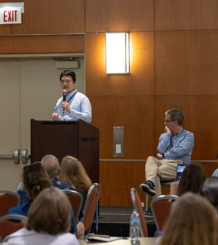
2026
PFIC Research Advancements Part 1 (Panel Presentations)
This panel brings together researchers at the forefront of PFIC science to share the latest developments in genetic diagnosis and emerging gene-targeted therapies — including a first look at a real patient case using a customized gene therapy approach.
Moderated by Dr. Chunyue Yin.
Featured speakers:
🔹 Dr. Chunyue Yin, Cincinnati Children’s
🔹 Dr. Richard Thompson — New Developments in Genetic Testing & Diagnosis
🔹 Dr. Henkjan Verkade — Disease Mechanisms & Gene Therapy Progress
🔹 Dr. Akihiro Asai, Cincinnati Children’s — Gene Therapy Deep Dive: The Case of Baby KJ
This session was recorded at the 2026 PFIC Family & Scientific Conference in Chicago, IL and sponsored by The Forum For Collaborative Research.
Did you like this video?
Whether you’re newly diagnosed or years into your PFIC journey, our webinar library has something for you. Browse topics like genetics, treatment, transplant, and coping strategies, all in one place.